Understanding Animal Cell Structures: Animal Cell Coloring Guide Answer Key
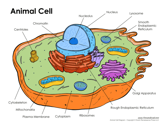
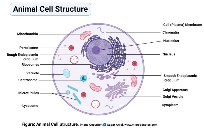
Animal cell coloring guide answer key – Animal cells are the fundamental units of life in animals, responsible for carrying out all essential biological processes. They are eukaryotic cells, meaning they have a defined nucleus and other membrane-bound organelles, each performing a specific function to maintain the cell’s life and contribute to the overall functioning of the organism.The primary components of a typical animal cell include the cell membrane, cytoplasm, nucleus, and various organelles such as mitochondria, ribosomes, endoplasmic reticulum, Golgi apparatus, lysosomes, and vacuoles.
These components work together in a coordinated manner to ensure the cell’s survival and proper functioning.
Working with an animal cell coloring guide answer key can be a great learning tool. For a fun break and to explore more visually appealing animal depictions, check out these cute coloring page animals. Then, return to your animal cell diagram to solidify your understanding of organelles and their functions.
Cell Membrane
The cell membrane, also known as the plasma membrane, is a selectively permeable barrier that surrounds the cell, separating its internal environment from the external surroundings. It regulates the passage of substances into and out of the cell, maintaining a stable internal environment. The cell membrane is primarily composed of a phospholipid bilayer with embedded proteins.
Cytoplasm
The cytoplasm is the jelly-like substance that fills the cell’s interior, encompassing all the organelles except the nucleus. It provides a medium for various cellular processes to occur and houses the cytoskeleton, a network of protein filaments that provides structural support and facilitates cell movement.
Nucleus
The nucleus is the control center of the cell, containing the cell’s genetic material in the form of DNA organized into chromosomes. The nucleus regulates gene expression and controls cell division. It is enclosed by a double membrane called the nuclear envelope, which contains pores that allow the transport of molecules between the nucleus and the cytoplasm.
Mitochondria
Mitochondria are often referred to as the “powerhouses” of the cell because they are responsible for generating energy through cellular respiration. They convert nutrients into ATP (adenosine triphosphate), the cell’s primary energy currency.
Ribosomes
Ribosomes are responsible for protein synthesis. They are small structures composed of RNA and protein, found either free in the cytoplasm or attached to the endoplasmic reticulum.
Endoplasmic Reticulum
The endoplasmic reticulum (ER) is a network of membranes involved in protein synthesis, lipid metabolism, and calcium storage. There are two types of ER: rough ER, which is studded with ribosomes and involved in protein synthesis, and smooth ER, which lacks ribosomes and is involved in lipid synthesis and detoxification.
Golgi Apparatus
The Golgi apparatus, also known as the Golgi complex, is responsible for modifying, sorting, and packaging proteins and lipids for transport to their final destinations within or outside the cell.
Lysosomes
Lysosomes are membrane-bound organelles containing digestive enzymes that break down waste materials, cellular debris, and foreign substances. They act as the cell’s recycling and waste disposal system.
Vacuoles
Vacuoles are membrane-bound sacs involved in storage, transport, and maintaining cell turgor pressure. Animal cells typically have smaller vacuoles compared to plant cells.
Comparison of Plant and Animal Cells
Plant and animal cells share many similarities, but they also have key structural differences. Plant cells have a cell wall made of cellulose, providing rigidity and support, while animal cells lack a cell wall. Plant cells also contain chloroplasts, the site of photosynthesis, which are absent in animal cells. Additionally, plant cells typically have a large central vacuole that plays a crucial role in maintaining cell turgor pressure, while animal cells have smaller vacuoles or may lack them altogether.
A table summarizing these differences is shown below:
| Feature | Plant Cell | Animal Cell |
|---|---|---|
| Cell Wall | Present | Absent |
| Chloroplasts | Present | Absent |
| Central Vacuole | Large | Small or absent |
Educational Applications of Animal Cell Diagrams

Animal cell diagrams and coloring guides serve as valuable tools for visualizing and understanding the complex structures within animal cells. They transform abstract concepts into tangible representations, fostering deeper learning and engagement in biological studies. The interactive nature of coloring encourages active participation, promoting knowledge retention and comprehension of cell structure and function.
Visualizing Cell Structures
Coloring guides enhance the learning process by allowing students to visualize the different organelles within an animal cell. Assigning specific colors to each organelle helps students differentiate them and understand their unique roles. For example, coloring the nucleus purple, the mitochondria orange, and the endoplasmic reticulum blue provides a visual representation that reinforces the spatial organization and distinct identities of these components.
This visual learning approach strengthens comprehension and memory recall of cell structure.
Assessment of Understanding
Coloring guides can be effectively used to assess student understanding of animal cell structure and function. By evaluating the accuracy of the coloring and accompanying labels, educators can gauge students’ grasp of organelle identification and their respective functions. For instance, if a student correctly colors and labels the Golgi apparatus, it demonstrates their understanding of its role in protein processing and packaging.
Furthermore, open-ended questions related to the coloring activity, such as describing the function of a specific organelle or explaining the relationship between two organelles, can provide deeper insights into student comprehension.
Integration into Lesson Plans
Animal cell diagrams can be seamlessly integrated into lesson plans across different grade levels.
- Elementary School: Introduce basic organelles like the nucleus, cytoplasm, and cell membrane using simplified diagrams and coloring activities. Focus on the general concept of cells as the building blocks of life.
- Middle School: Explore a wider range of organelles, including the mitochondria, ribosomes, and endoplasmic reticulum. Incorporate interactive coloring guides that challenge students to label and describe the function of each organelle.
- High School: Delve deeper into the complexities of cellular processes, such as protein synthesis, cellular respiration, and cell division. Utilize detailed diagrams and coloring guides to illustrate these intricate processes and their connection to the various organelles.
For example, in a high school biology lesson on protein synthesis, students can use a coloring guide to track the path of a protein from its creation in the ribosomes, through the endoplasmic reticulum and Golgi apparatus, to its final destination. This activity reinforces the interconnectedness of organelles and their roles in this essential cellular process.
Visualizing Animal Cells

Understanding the visual characteristics of animal cells is crucial for accurate representation and interpretation of their structure and function. This section provides detailed descriptions of key organelles and their appearance, offering a comprehensive guide for visualizing and illustrating animal cells.
Appearance and Structure of Key Organelles
The nucleus, often centrally located, appears as a large, spherical or ovoid body. It houses the cell’s genetic material (DNA) and is typically darker staining than the surrounding cytoplasm due to its dense content. The nuclear envelope, a double membrane, surrounds the nucleus, punctuated by nuclear pores that regulate the passage of molecules. Within the nucleus, the nucleolus, a dense, spherical structure, is the site of ribosome production.Mitochondria, the powerhouses of the cell, are rod-shaped or oval organelles with a double membrane.
The inner membrane is highly folded into cristae, which increase the surface area for cellular respiration. Under a microscope, mitochondria may appear as elongated, granular structures scattered throughout the cytoplasm.The endoplasmic reticulum (ER) is a network of interconnected membranes forming flattened sacs and tubules. Rough ER is studded with ribosomes, giving it a granular appearance, and is involved in protein synthesis.
Smooth ER lacks ribosomes and appears smoother under a microscope. It plays a role in lipid synthesis and detoxification.Ribosomes, the protein synthesis machinery, are small, granular structures found free in the cytoplasm or attached to the rough ER. They are too small to visualize detailed structure with a light microscope, appearing as tiny dots.The Golgi apparatus, involved in processing and packaging proteins, appears as a stack of flattened, membrane-bound sacs, or cisternae.
It is often located near the nucleus and can be visualized as a distinct, slightly curved structure.Lysosomes, responsible for cellular digestion, are small, spherical organelles containing digestive enzymes. They appear as dark, granular bodies under a microscope.The cell membrane, enclosing the entire cell, is a thin, flexible barrier that regulates the passage of substances into and out of the cell.
It is not easily visible with a light microscope but defines the cell’s boundary.
Microscopic View of an Animal Cell
Under a light microscope, an animal cell appears as a three-dimensional structure bounded by a cell membrane. The cytoplasm, a clear, jelly-like substance, fills the cell and contains various organelles. The nucleus, often the most prominent organelle, is typically centrally located and appears darker than the cytoplasm. Scattered throughout the cytoplasm are smaller organelles such as mitochondria, which may appear as elongated granules, and the Golgi apparatus, visible as a stacked structure.
Ribosomes and lysosomes are generally too small to resolve detailed structure with a light microscope, appearing as tiny dots or granules.
Organelle Size Organization, Animal cell coloring guide answer key
An introductory explanation about organelle sizes and their respective functions in the cell is important for understanding the cell’s internal organization. The size of an organelle can often relate to its role and the volume of its activity within the cell.
| Organelle | Size Range (approximate) | Description |
|---|---|---|
| Nucleus | 5-10 μm | Contains the cell’s genetic material and controls cell activities. |
| Mitochondria | 0.5-10 μm | Generates energy for the cell through cellular respiration. |
| Golgi Apparatus | 1-3 μm | Processes, packages, and distributes proteins and lipids. |
| Endoplasmic Reticulum (ER) | Variable, network structure | Synthesizes proteins and lipids, and detoxifies harmful substances. |
| Lysosomes | 0.1-1.2 μm | Digest cellular waste and foreign materials. |
| Ribosomes | ~20 nm | Synthesize proteins. |
