Animal Cell Structure and Components
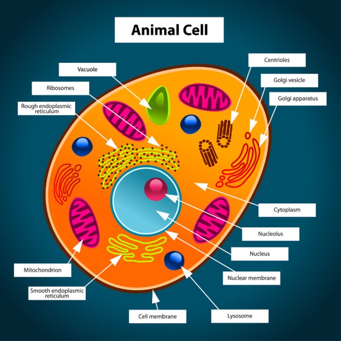
Animal cell coloring answers – Animal cells, the fundamental building blocks of animals, are complex structures teeming with specialized organelles, each performing a unique role to maintain the cell’s life and function. Understanding these components is crucial to grasping the intricacies of animal biology. This section delves into the key organelles and their roles, exploring the cell membrane’s vital function and highlighting the differences between animal and plant cells.
Major Organelles and Their Functions
The animal cell is a bustling metropolis of organelles, each with a specific job to ensure the smooth operation of the cell. These organelles work together in a coordinated manner to maintain cellular homeostasis and carry out essential life processes.
| Organelle | Function | Description | Example |
|---|---|---|---|
| Nucleus | Contains genetic material (DNA) and controls cell activities. | The control center of the cell, dictating protein synthesis and cell division. | The nucleus directs the production of enzymes needed for cellular respiration. |
| Ribosomes | Synthesize proteins. | Tiny structures found free-floating in the cytoplasm or attached to the endoplasmic reticulum. | Ribosomes in the pancreas produce insulin. |
| Endoplasmic Reticulum (ER) | Synthesizes lipids and proteins; transports materials within the cell. | A network of membranes extending throughout the cytoplasm; rough ER (with ribosomes) synthesizes proteins, smooth ER synthesizes lipids. | The smooth ER in liver cells helps detoxify harmful substances. |
| Golgi Apparatus | Modifies, sorts, and packages proteins and lipids for secretion or use within the cell. | A stack of flattened sacs that acts as a processing and distribution center. | The Golgi apparatus packages digestive enzymes for lysosomes. |
| Mitochondria | Generate energy (ATP) through cellular respiration. | The “powerhouses” of the cell, converting nutrients into usable energy. | Muscle cells have many mitochondria to fuel their contractions. |
| Lysosomes | Digest waste materials and cellular debris. | Membrane-bound sacs containing digestive enzymes. | Lysosomes break down old or damaged organelles. |
| Cytoskeleton | Provides structural support and facilitates cell movement. | A network of protein filaments (microtubules, microfilaments, intermediate filaments) that maintains cell shape and aids in intracellular transport. | The cytoskeleton enables cell division and cell migration. |
| Cell Membrane | Regulates the passage of substances into and out of the cell. | A selectively permeable barrier that encloses the cell’s contents. | The cell membrane controls the flow of ions like sodium and potassium. |
Cell Membrane Structure and Homeostasis
The cell membrane is a fluid mosaic of phospholipids and proteins, acting as a selective barrier that controls the movement of substances in and out of the cell. This selective permeability is crucial for maintaining homeostasis, the cell’s internal balance. The phospholipid bilayer, with its hydrophobic tails and hydrophilic heads, forms the basic structure, while embedded proteins facilitate transport, cell signaling, and other functions.
The membrane’s fluidity allows for dynamic changes in its composition and function, adapting to the cell’s needs. For example, the sodium-potassium pump, an integral membrane protein, actively transports sodium ions out of the cell and potassium ions into the cell, maintaining the cell’s electrochemical gradient.
Differences Between Plant and Animal Cells
While both plant and animal cells are eukaryotic, possessing membrane-bound organelles, several key differences exist. Plant cells possess a rigid cell wall made of cellulose, providing structural support and protection, which is absent in animal cells. Plant cells also contain chloroplasts, the sites of photosynthesis, enabling them to produce their own food, a process not performed by animal cells.
Finally, plant cells typically have a large central vacuole for storage and maintaining turgor pressure, while animal cells may have smaller vacuoles or lack them altogether. These differences reflect the distinct lifestyles and functions of plant and animal organisms.
Cell Coloring Activities: Animal Cell Coloring Answers

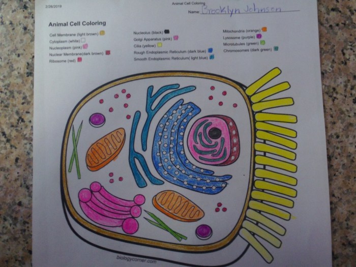
Bringing your animal cell diagrams to life through coloring isn’t just a fun activity; it’s a powerful learning tool. By assigning specific colors to different organelles, you create a visual map that reinforces understanding of their structure and function. This process enhances memory retention and allows for a deeper comprehension of the intricate workings within an animal cell.Coloring methods offer diverse approaches to this educational endeavor.
Whether you prefer the tactile experience of traditional media or the precision of digital tools, the key is accuracy and thoughtful color selection.
Methods and Techniques for Coloring Animal Cell Diagrams
Crayons, colored pencils, and digital painting software each provide unique advantages for coloring animal cell diagrams. Crayons offer vibrant, bold colors and are ideal for younger learners due to their ease of use. However, achieving fine detail can be challenging. Colored pencils, on the other hand, allow for more nuanced shading and detail, enabling a more realistic representation of the cell’s structures.
Digital tools, such as Adobe Photoshop or Procreate, offer the greatest flexibility, allowing for intricate details, layering, and easy corrections. The choice of medium depends largely on personal preference and the desired level of detail.
Understanding animal cell coloring answers requires a grasp of cellular structures. For a fun, visual aid before tackling those diagrams, check out these adorable printable coloring sheets animals – they’re a great way to relax and appreciate the beauty of the animal kingdom, which can inspire a deeper understanding of animal cell components later. Returning to the cellular level, remember to accurately color organelles like the nucleus and mitochondria for complete animal cell coloring answers.
Step-by-Step Procedure for Accurate Cell Diagram Coloring
Let’s assume we’re working with a diagram showing a typical animal cell, including the nucleus, mitochondria, endoplasmic reticulum, Golgi apparatus, ribosomes, lysosomes, and cell membrane.
1. Preparation
Begin by having a clear, labeled diagram of an animal cell. Choose your coloring medium (crayons, colored pencils, or digital tools).
2. Nucleus
Color the nucleus a dark purple or deep blue to represent its role as the control center containing genetic material. This rich color helps it stand out as a key component.
3. Mitochondria
Depict the mitochondria in a reddish-brown or burnt orange. This color choice symbolizes their energy-producing function, reminiscent of fire or heat. Draw them as elongated oval shapes scattered throughout the cytoplasm.
4. Endoplasmic Reticulum
Color the endoplasmic reticulum (ER) a light blue or pale green. This softer color reflects its role in protein synthesis and transport. Illustrate the ER as a network of interconnected membranes, both rough (studded with ribosomes) and smooth.
5. Golgi Apparatus
Represent the Golgi apparatus in a light yellow or beige. This color suggests the processing and packaging function of this organelle. Show it as a stack of flattened sacs near the nucleus.
6. Ribosomes
Since ribosomes are small, use tiny dots of dark purple or dark blue to represent them, scattered throughout the cytoplasm and attached to the rough ER.
7. Lysosomes
Color the lysosomes a light orange or pinkish-red, suggesting their role in waste breakdown. These should be smaller, rounded structures scattered throughout the cell.
8. Cell Membrane
Use a thin line of dark brown or black to Artikel the cell membrane, highlighting its boundary role.
Color-Coding Schemes for Organelles
Choosing a color-coding scheme for organelles isn’t arbitrary; it should reflect their functions and enhance visual understanding.A suggested scheme:
- Nucleus: Deep blue/purple (control center, DNA storage)
- Mitochondria: Reddish-brown/Burnt orange (energy production)
- Endoplasmic Reticulum: Light blue/Pale green (protein synthesis & transport)
- Golgi Apparatus: Light yellow/Beige (processing & packaging)
- Ribosomes: Dark purple/Dark blue (protein synthesis)
- Lysosomes: Light orange/Pinkish-red (waste breakdown)
- Cell Membrane: Dark brown/Black (boundary & protection)
The rationale behind these choices is to use colors that visually associate with the organelle’s function. For instance, the reddish-brown of mitochondria evokes the idea of heat and energy production, while the pale green of the ER suggests growth and synthesis. This color association aids in memory retention and a deeper understanding of cellular processes.
Interpreting Animal Cell Coloring Exercises

Accurately coloring an animal cell diagram isn’t just a fun activity; it’s a crucial step in understanding the intricate workings of this fundamental unit of life. The process of carefully identifying and coloring each organelle forces a deeper engagement with the cell’s structure and, consequently, its function. This active learning approach enhances retention and provides a visual framework for understanding complex biological processes.Accurate coloring directly relates to understanding cell function.
Each organelle has a specific role, and its visual representation – its color and location within the cell – should reflect this. For instance, the vibrant green of a chloroplast (though absent in animal cells) immediately signals its role in photosynthesis. Similarly, the distinct coloring of the nucleus highlights its crucial role as the cell’s control center, housing the genetic material.
Misrepresenting these organelles through inaccurate coloring can lead to a flawed understanding of their individual functions and their interplay within the cell.
Identifying Errors in Incorrectly Colored Animal Cell Diagrams
Consider a hypothetical scenario: an animal cell diagram depicts the mitochondria as a pale blue, indistinguishable from the surrounding cytoplasm. This error undermines the understanding of the mitochondria’s critical role in cellular respiration, energy production, and the cell’s overall metabolism. Another common mistake is misplacing organelles. For example, if the Golgi apparatus is depicted inside the nucleus, it misrepresents its role in protein modification and transport, which occurs outside the nucleus.
Incorrect size ratios between organelles also represent a significant error. The nucleus, for example, should be proportionally larger than the ribosomes. These visual inaccuracies create a misleading representation of the cell’s structure and function.
Rubric for Evaluating Animal Cell Coloring Exercises
A comprehensive rubric for evaluating an animal cell coloring exercise should consider several key aspects. It should assess not only the accuracy of the coloring but also the completeness of the diagram. Here’s a possible rubric:
| Criteria | Excellent (4 points) | Good (3 points) | Fair (2 points) | Poor (1 point) |
|---|---|---|---|---|
| Accuracy of Organelle Identification | All organelles correctly identified and colored. | Most organelles correctly identified and colored; minor inaccuracies. | Several organelles incorrectly identified or colored. | Many organelles incorrectly identified or colored; significant inaccuracies. |
| Accuracy of Organelle Location | All organelles correctly positioned within the cell. | Most organelles correctly positioned; minor inaccuracies in placement. | Several organelles incorrectly positioned. | Many organelles incorrectly positioned; significant inaccuracies. |
| Accuracy of Organelle Size Ratio | Accurate representation of relative sizes of organelles. | Mostly accurate representation of relative sizes; minor discrepancies. | Significant discrepancies in relative sizes of organelles. | Inaccurate representation of relative sizes; major discrepancies. |
| Neatness and Presentation | Diagram is neat, well-organized, and clearly labeled. | Diagram is mostly neat and well-organized; minor labeling issues. | Diagram is somewhat disorganized; some labeling issues. | Diagram is messy and poorly organized; significant labeling issues. |
Visual Representation of Cell Processes Through Coloring
Different cell processes can be visually represented through coloring by emphasizing specific organelles involved. For example, the process of protein synthesis can be highlighted by using a bright color for ribosomes (where protein synthesis begins) and a different color for the endoplasmic reticulum and Golgi apparatus (where proteins are modified and transported). Similarly, the process of cellular respiration can be visualized by using a distinct color for the mitochondria, emphasizing their role in energy production.
This approach helps students connect the visual representation with the underlying biological processes. The contrast in colors helps emphasize the dynamic interplay between different organelles in carrying out these complex cellular functions.
Advanced Concepts in Animal Cell Structure and Function

Delving deeper into the intricacies of animal cells reveals a fascinating world of complex processes and interactions. Understanding these advanced concepts is crucial for comprehending the overall function and health of an organism. This section explores key cellular processes, focusing on the mitochondria, endoplasmic reticulum, and Golgi apparatus.
Cellular Respiration in Mitochondria, Animal cell coloring answers
Mitochondria, often dubbed the “powerhouses” of the cell, are responsible for cellular respiration, the process that converts nutrients into usable energy in the form of ATP (adenosine triphosphate). This process occurs in several stages. First, glycolysis breaks down glucose in the cytoplasm, producing pyruvate. Pyruvate then enters the mitochondria, where it undergoes oxidative decarboxylation, converting it into acetyl-CoA.
The citric acid cycle (Krebs cycle) follows, a series of reactions that further oxidize acetyl-CoA, releasing carbon dioxide and generating high-energy electron carriers (NADH and FADH2). Finally, these electron carriers donate their electrons to the electron transport chain, a series of protein complexes embedded in the inner mitochondrial membrane. As electrons move down the chain, energy is released and used to pump protons (H+) across the membrane, creating a proton gradient.
This gradient drives ATP synthesis through chemiosmosis, where protons flow back across the membrane through ATP synthase, an enzyme that phosphorylates ADP to ATP. The final electron acceptor is oxygen, which combines with protons to form water. This intricate process ensures a continuous supply of energy for cellular activities.
Endoplasmic Reticulum and Protein Synthesis
The endoplasmic reticulum (ER) plays a vital role in protein synthesis and transport. The ER is a network of interconnected membranes extending throughout the cytoplasm. Rough ER, studded with ribosomes, is the primary site of protein synthesis. Ribosomes translate mRNA into polypeptide chains, which then enter the lumen of the ER for folding and modification. Smooth ER, lacking ribosomes, is involved in lipid synthesis, detoxification, and calcium storage.
Proteins synthesized on the rough ER are often destined for secretion or incorporation into membranes. These proteins undergo various modifications within the ER lumen, including glycosylation (addition of sugar molecules) and disulfide bond formation, ensuring proper folding and function. After modification, proteins are packaged into transport vesicles for delivery to the Golgi apparatus.
Golgi Apparatus and Cell Secretion
The Golgi apparatus, a stack of flattened membrane-bound sacs (cisternae), acts as a processing and packaging center for proteins and lipids. Proteins arriving from the ER enter the cis face (receiving side) of the Golgi, where they undergo further modifications, such as glycosylation and proteolytic cleavage. As proteins move through the Golgi cisternae, they are sorted and packaged into vesicles based on their destination.
The trans face (shipping side) of the Golgi buds off vesicles containing processed proteins destined for secretion, lysosomes, or other cellular compartments. This precise sorting mechanism ensures that proteins reach their correct locations within the cell or are secreted outside the cell. The Golgi apparatus is crucial for cell secretion, enabling the release of hormones, enzymes, and other molecules vital for cellular communication and function.
Protein Synthesis: A Step-by-Step Overview
The synthesis of a protein from DNA to its secretion involves a series of intricate steps:* Transcription: The DNA sequence of a gene is transcribed into a messenger RNA (mRNA) molecule.
mRNA Processing
The pre-mRNA undergoes processing, including splicing (removal of introns) and addition of a 5′ cap and a 3′ poly(A) tail.
Translation
The processed mRNA molecule binds to a ribosome, and the sequence is translated into a polypeptide chain.
Protein Folding
The polypeptide chain folds into its three-dimensional structure, often assisted by chaperone proteins.
Post-translational Modification
The protein undergoes modifications, such as glycosylation or phosphorylation, in the ER and Golgi apparatus.
Packaging and Transport
The modified protein is packaged into transport vesicles and transported to its final destination, either within the cell or secreted outside the cell.
Secretion
Secretory proteins are released from the cell through exocytosis.
Quick FAQs
What are some common mistakes made when coloring animal cells?
Common mistakes include inaccurate sizing and placement of organelles, inconsistent color choices, and neglecting to label structures clearly.
How can I adapt animal cell coloring for different learning abilities?
Adaptations include using larger diagrams for visual learners, providing simplified versions for younger students, and offering tactile options like textured paper for kinesthetic learners.
Are there online resources to help with animal cell coloring?
Yes! Many websites and educational platforms offer printable diagrams, interactive activities, and virtual coloring tools for animal cells.
What are the benefits of using color-coding in animal cell diagrams?
Color-coding improves visual organization, clarifies relationships between organelles, and aids memory retention of structure and function.
