Creating a Visual Guide

Animal cell coloring guide – Yo, Medan peeps! Let’s get this cell diagram lookingsick*. We’re talking about making an animal cell illustration that’s not just informative, but also totally eye-catching. Think Instagram-worthy, but for science class.Creating a killer visual guide for an animal cell needs the right approach. You can go old-school with a hand-drawn masterpiece or jump into the digital world for some seriously polished results.
Both have their own vibes, and we’ll break down the pros and cons of each.
Hand-Drawn Methods
Hand-drawing your animal cell offers a unique, personal touch. Think of it as your own artistic interpretation of cellular biology. Using colored pencils, markers, or even watercolors, you can create a really expressive diagram. The downside? It can be time-consuming and achieving perfect accuracy can be a challenge.
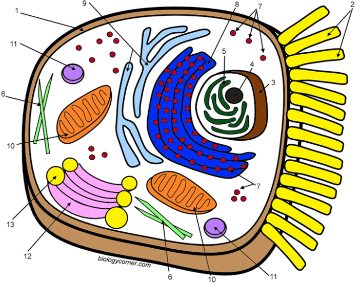
Getting the proportions right for all the organelles takes some serious skill and patience. A good hand-drawn diagram will use bold Artikels to clearly define each organelle and strategically use color to differentiate between structures. For example, the nucleus could be a deep purple, the mitochondria a vibrant red, and the ribosomes a speckled light blue.
Digital Methods
Digital tools like Adobe Illustrator or Procreate offer incredible precision and editing capabilities. You can easily adjust sizes, colors, and add intricate details. This gives you way more control over the final product and allows for cleaner, more accurate representations. The drawback? It requires familiarity with the software.
A beginner might find it overwhelming at first. However, the learning curve is worth it for the level of detail and professional look you can achieve. A well-executed digital illustration might use gradients to create a three-dimensional effect, giving the cell a more realistic appearance.
Effective Visual Representations, Animal cell coloring guide
Think about textbook diagrams of animal cells. Some show a simplified, almost cartoonish representation, focusing on the major organelles. These are great for quick understanding, but they lack detail. Others are incredibly detailed, almost microscopic in their accuracy. These can be overwhelming for beginners but excellent for those who need to delve into the specifics.
The best visual representations find a balance between clarity and detail, using clear labeling and color-coding to make complex structures easy to understand.
Shading and Texture
Adding shading and texture is where your diagram goes from “meh” to “wow.” Shading gives organelles depth and dimension, making them appear three-dimensional. For example, you could use a light source to create shadows on one side of the nucleus, giving it a round, plump look. Texture can be added to represent the different components. For instance, the rough endoplasmic reticulum could be depicted with a slightly rough texture, contrasting with the smooth endoplasmic reticulum’s smoother appearance.
This helps to emphasize the differences in their functions and structures. Using a combination of these techniques makes your diagram visually engaging and easier to comprehend.
Illustrative Examples of Animal Cell Components: Animal Cell Coloring Guide

Yo, Medan peeps! Let’s dive into the wild world of animal cells. We’re gonna break down the key players – the organelles – and how to spot ’em like a pro. Think of this as your ultimate cheat sheet for acing that cell biology assignment (or just impressing your friends).
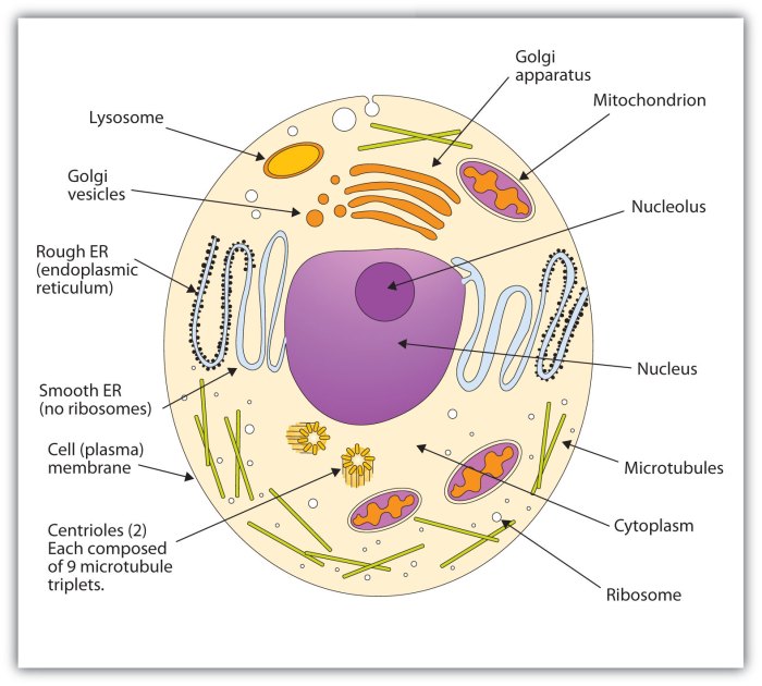
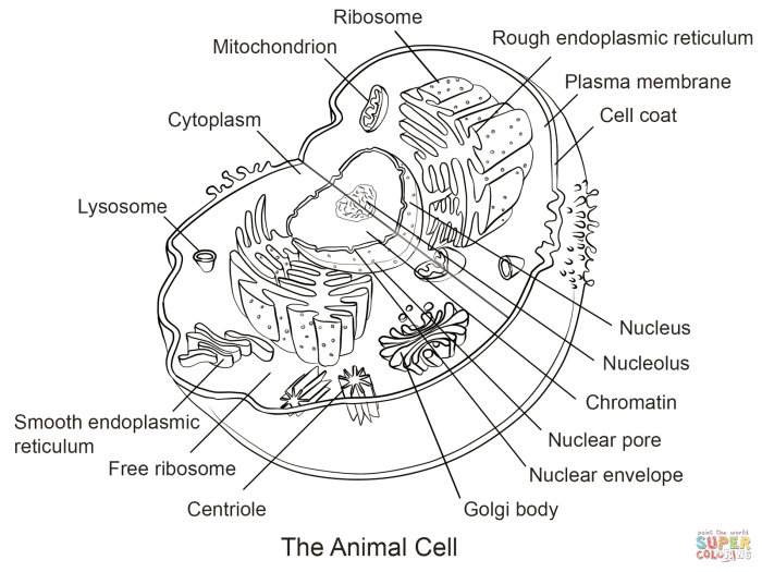
Understanding the unique visual characteristics of each organelle is crucial for accurate identification. We’ll be looking at size, shape, internal structure, and location within the cell to help you tell them apart. Think of it as a cellular fashion show, where each organelle rocks its own unique style!
Nucleus
The nucleus is the undisputed boss of the cell, kinda like the mayor of Cell City. It’s usually the biggest and most noticeable organelle, a round or oval-shaped structure hanging out near the center. Imagine a large, perfectly spherical bubble, and that’s kinda the gist of it. You’ll see a distinct boundary – the nuclear envelope – surrounding it.
Inside, you might be able to spot the nucleolus, a slightly darker region responsible for making ribosomes. Think of it as the nucleus’s super-busy workshop.
Mitochondria
These guys are the powerhouses, the energy factories of the cell. They’re usually sausage-shaped or oval, with a slightly wrinkled outer membrane and a highly folded inner membrane. This folded inner membrane is key – it’s called cristae, and it significantly increases the surface area for energy production. Imagine a tiny, wrinkled bean, but way cooler.
Ribosomes
Ribosomes are the tiny protein-making machines. They’re super small, so you might need a really powerful microscope to see them clearly. They appear as tiny dots, either freely floating in the cytoplasm or attached to the endoplasmic reticulum. Think of them as millions of tiny, industrious workers building the cell’s proteins.
Endoplasmic Reticulum
The endoplasmic reticulum (ER) is a network of interconnected membranes, like a vast highway system within the cell. There are two types: rough ER (studded with ribosomes, giving it a bumpy appearance) and smooth ER (lacking ribosomes, appearing as a smooth network of tubules). The rough ER looks like a bunch of interconnected, bumpy sacs, while the smooth ER looks more like a system of smooth, interconnected tubes.
Golgi Apparatus
The Golgi apparatus, also known as the Golgi body or Golgi complex, is like the cell’s packaging and shipping department. It’s usually a stack of flattened, membrane-bound sacs (cisternae). Imagine a stack of pancakes, but made of membranes. It modifies, sorts, and packages proteins and lipids for transport to other parts of the cell or outside the cell.
Lysosomes
Lysosomes are the cell’s recycling and waste disposal system. They are small, membrane-bound sacs containing digestive enzymes. They appear as small, round vesicles scattered throughout the cytoplasm. Think of them as the cell’s cleanup crew, breaking down waste materials and cellular debris.
Vacuoles
Vacuoles are membrane-bound sacs that store various substances, such as water, nutrients, or waste products. In animal cells, they are generally smaller and more numerous than in plant cells. They can vary in size and shape, but they generally appear as small, clear sacs within the cytoplasm. Think of them as the cell’s storage containers.
Simple Diagram of Organelle Size and Location
Imagine a cell as a bustling city. The nucleus is the city hall, large and central. The mitochondria are like the power plants scattered throughout, providing energy. The endoplasmic reticulum is the highway system, connecting different parts of the city. The Golgi apparatus is the post office, packaging and shipping goods.
Lysosomes are the sanitation department, cleaning up waste. Ribosomes are the tiny construction workers building everywhere. Finally, vacuoles are the storage facilities holding various materials.
Understanding animal cell structure is key to biology education, and a helpful visual aid is an animal cell coloring guide. For a fun, related activity, especially for younger learners, consider supplementing this with some engaging visuals, like the adorable farm animals printable coloring pages , which can help reinforce the concept of different organisms and their cellular structures.
Returning to the animal cell coloring guide, remember to carefully label each organelle for a complete learning experience.
Advanced Techniques

Level up your animal cell game, Medan style! We’ve covered the basics, now let’s dive into some seriously cool techniques to make your cell diagrams pop. Think beyond flat drawings – we’re talking 3D models and interactive guides that’ll make learning cell biology a breeze.
3D Modeling Advantages and Challenges
Creating a 3D model of an animal cell offers a fantastic way to visualize its complex structure and the spatial relationships between its organelles. Imagine a mini-universe within a cell, brought to life! However, building a convincing 3D model isn’t a walk in the park. It requires specialized software, a good understanding of cell biology, and a fair bit of patience.
The advantages include improved understanding of spatial relationships, enhanced engagement for learners, and a more memorable learning experience. The challenges include the complexity of modeling intricate structures, the need for specialized software and skills, and the potential for inaccuracies if not based on reliable sources. Think of it like building a super detailed Lego castle – awesome, but also time-consuming!
Creating an Interactive Animal Cell Diagram
Let’s craft a simple interactive animal cell diagram using readily available tools like PowerPoint or Google Slides. This is a super accessible method to create something engaging, even without fancy 3D modeling software.
-
Step 1: Layout and Organelles: Start with a basic circle representing the cell membrane. Then, add shapes representing the major organelles (nucleus, mitochondria, ribosomes, endoplasmic reticulum, Golgi apparatus, lysosomes, etc.). Use different colors and fill styles to differentiate them. Think of it as arranging your favorite Medan street food – each item has its own unique color and shape!
-
Step 2: Labeling and Annotation: Add text boxes to label each organelle. You can use simple labels or more descriptive ones, depending on the complexity you want. Consider using a consistent font and size for a clean look. Imagine it’s like labeling the ingredients on your favorite Nasi Lemak – clear, concise, and easy to understand!
-
Step 3: Adding Interactivity (PowerPoint): PowerPoint offers animation features. You can create simple animations to highlight each organelle upon clicking. For example, when you click on the nucleus, it could slightly enlarge or glow. This is like adding a little surprise to your favorite dish – a wow factor!
-
Step 4: Adding Interactivity (Google Slides): Google Slides offers similar functionality to PowerPoint, allowing for animations and transitions. You can also embed links to external resources within the diagram, linking each organelle to more information online. It’s like adding extra notes to your recipe, pointing to where you got the inspiration!
-
Step 5: Review and Refinement: Once you’ve added all the organelles and interactivity, take a step back and review your work. Make sure everything is clearly labeled, the colors are distinct, and the animations are smooth and easy to follow. It’s like tasting your dish before serving – making sure everything is perfect!
Quick FAQs
What are the best digital tools for creating interactive animal cell diagrams?
PowerPoint, Google Slides, and specialized biology software offer excellent interactive capabilities. Choose the tool that best suits your skill level and desired level of interactivity.
How can I adapt this guide for different age groups?
Simplify the descriptions and color-coding for younger learners. For older students, incorporate more complex details and advanced techniques like 3D modeling.
Are there any readily available pre-made animal cell coloring pages?
Yes, many websites and educational resources offer printable animal cell coloring pages. However, creating your own allows for greater customization and deeper understanding.
What are some alternative color schemes for animal cell organelles?
Experiment with different palettes! Consider using analogous colors for related organelles or complementary colors for contrasting structures. The key is clear visual distinction.
