Target Audience for Animal Cell Coloring Pages
Animal cell coloring pages serve as a valuable educational tool for visualizing and understanding the complex structure of animal cells. They cater to a diverse audience, primarily focused on learning and exploring biology, but also extending to individuals seeking creative outlets or engaging activities.The primary demographic for animal cell coloring pages includes students, educators, and homeschooling families. These individuals utilize coloring pages as a supplementary learning resource to enhance understanding of cellular biology.
Beyond the educational sphere, individuals interested in science, art, or creative expression may also find animal cell coloring pages appealing.
Age Groups and Their Needs
Different age groups benefit from animal cell coloring pages tailored to their specific learning levels and cognitive abilities.
- Elementary School (Ages 6-10): Simplified diagrams focusing on basic organelles like the nucleus, cytoplasm, and cell membrane are ideal for this age group. Large, clearly defined areas for coloring facilitate fine motor skill development.
- Middle School (Ages 11-13): More detailed diagrams incorporating additional organelles like mitochondria, ribosomes, and the endoplasmic reticulum are appropriate. Activities that connect organelle function to coloring can enhance comprehension.
- High School (Ages 14-18): Intricate diagrams depicting the complex interplay of organelles and cellular processes are suitable for this age group. Coloring can be integrated with labeling exercises and advanced biological concepts.
Educational Levels
Animal cell coloring pages are beneficial across various educational levels, from elementary school to higher education.
- K-12 Education: Coloring pages provide a hands-on approach to learning about cell structure and function, making abstract concepts more tangible. They can be incorporated into lesson plans, homework assignments, or independent study activities.
- Higher Education: While less common, coloring pages can be used in introductory biology courses at the college level to review fundamental concepts or as a starting point for more in-depth exploration.
Educational Value of Animal Cell Coloring Pages

Animal cell coloring pages offer a dynamic and engaging approach to learning about cell biology. They transform the complex world of cellular structures into a visually accessible and interactive experience, benefiting learners of all ages. By actively participating in coloring, students move beyond passive memorization and develop a deeper understanding of the intricate components and functions of animal cells.Coloring pages bridge the gap between abstract concepts and tangible representations.
The act of coloring encourages focused observation of each organelle’s shape, size, and position within the cell. This active engagement enhances comprehension and promotes a more meaningful learning experience compared to simply reading text or viewing diagrams.
Visual Learning and Memory Retention
Visual learning plays a crucial role in understanding complex scientific concepts. Coloring pages leverage this by transforming abstract cellular structures into concrete visual representations. The process of selecting colors and meticulously filling in the designated areas for each organelle reinforces visual memory. This active engagement strengthens the association between the visual representation and the organelle’s name and function.
Enhanced Understanding of Cell Biology Concepts, Animal cell coloring page
Animal cell coloring pages serve as effective tools for illustrating key cell biology concepts. For example, coloring the mitochondria a vibrant red can highlight its role as the “powerhouse” of the cell, generating energy. Coloring the Golgi apparatus a contrasting color, perhaps purple, emphasizes its distinct function in processing and packaging proteins. This color-coding approach helps students differentiate between various organelles and understand their individual roles within the complex cellular environment.
Examples of Coloring Page Applications
Coloring pages can be integrated into various learning activities to reinforce understanding. For instance, a student could color the nucleus a deep blue after learning about its role in containing DNA. Another activity might involve coloring the ribosomes a bright green, emphasizing their protein synthesis function. A more advanced exercise could involve labeling each colored organelle with its corresponding function.
These activities promote active recall and solidify understanding of the interrelationships between different cellular components.
Common Features of Animal Cell Coloring Pages
Animal cell coloring pages serve as an engaging educational tool for exploring the intricate world of biology. These pages typically depict the fundamental components of a typical animal cell, allowing for a visual understanding of their structure and function. Coloring encourages active learning, promoting better retention of information compared to passive reading.
Exploring the intricate details of an animal cell coloring page can be a great educational activity. For a change of pace, you might enjoy browsing free coloring pages anime , offering a different style of artistic expression. Afterward, return to your animal cell diagram and test your knowledge by coloring the organelles accurately.
Key Organelles and Structures
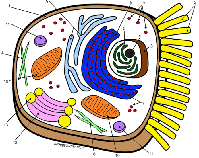
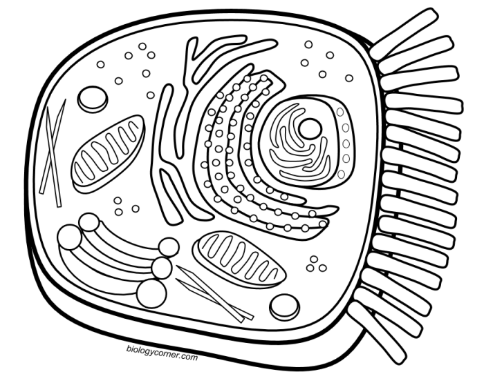
Animal cell coloring pages generally feature key organelles essential for cell function. These include the nucleus, mitochondria, ribosomes, endoplasmic reticulum (both smooth and rough), Golgi apparatus, lysosomes, and the cell membrane. Often, the cytoplasm is also represented, providing context for the organelles within the cell.
Description of an Appealing and Informative Animal Cell Coloring Page
An effective animal cell coloring page should be visually clear and informative. The cell membrane should be depicted as a distinct boundary, enclosing all other organelles. The nucleus, typically the largest organelle, should be centrally located and clearly labeled. Mitochondria, often depicted as bean-shaped structures, should be scattered throughout the cytoplasm. Ribosomes, smaller and more numerous, could be represented as dots or small circles.
The endoplasmic reticulum, a network of interconnected membranes, should be shown near the nucleus, with the rough ER distinguished by the presence of ribosomes on its surface. The Golgi apparatus, a stack of flattened sacs, should be positioned near the endoplasmic reticulum. Lysosomes, small spherical organelles, should be distributed throughout the cytoplasm. Clear labels for each organelle contribute to the educational value of the page.
The use of varied colors and textures can enhance visual appeal and make the learning experience more engaging. For example, the nucleus could be a light purple, the mitochondria a vibrant orange, and the endoplasmic reticulum a light blue.
Animal Cell Coloring Page with Labels
The following table provides a simplified representation of an animal cell coloring page suitable for labeling. Imagine each cell as a distinct area on the coloring page, with the organelle name serving as the label. This table-based layout can be easily adapted to a visual coloring page format.
| Nucleus | Mitochondria | Ribosomes | Endoplasmic Reticulum (Smooth) |
| Endoplasmic Reticulum (Rough) | Golgi Apparatus | Lysosomes | Cell Membrane |
| Cytoplasm |
Creative Uses for Animal Cell Coloring Pages
Animal cell coloring pages offer a dynamic and engaging approach to learning about cell biology. Beyond simply coloring within the lines, these pages can be transformed into interactive learning tools that foster a deeper understanding of cellular structures and functions. This section explores innovative ways to utilize animal cell coloring pages to enhance the learning experience.
Interactive Learning Activities with Animal Cell Coloring Pages
Animal cell coloring pages can be integrated into various interactive learning activities to make learning about cell biology more engaging. These activities cater to different learning styles and can be adapted to suit various age groups.
- Create a 3D Cell Model: After coloring the cell components, students can cut them out and layer them to create a three-dimensional representation of an animal cell. This hands-on activity helps visualize the spatial arrangement of organelles within the cell.
- Cell Component Matching Game: Prepare separate cards with the names and functions of different cell organelles. Students can then match these cards to the corresponding colored sections on their animal cell coloring page.
- Design Your Own Organelle: Encourage students to invent a new organelle and draw it on their coloring page. They should also describe its function and how it interacts with other organelles, fostering creativity and critical thinking.
Lesson Plan: Exploring the Animal Cell
This lesson plan utilizes animal cell coloring pages and other resources to teach students about the structure and function of animal cells.
| Objective | Students will be able to identify and describe the functions of major animal cell organelles. |
|---|---|
| Materials | Animal cell coloring pages, colored pencils or markers, scissors, glue, chart paper, markers, index cards. |
| Procedure |
|
| Assessment | Observe student participation in discussions and evaluate the accuracy of their 3D cell models and organelle card matching. |
Illustrating Animal Cells for Coloring Pages

Creating accurate and engaging animal cell illustrations for coloring pages requires a balance of scientific accuracy and artistic appeal. The illustration must clearly depict the key organelles while remaining simple enough for coloring and engaging for the target audience. This involves careful consideration of the cell’s structure, the choice of art style, and the overall composition of the illustration.
Creating a visually appealing and scientifically accurate animal cell illustration for a coloring page involves several steps. First, research and gather reference materials depicting animal cell structure. This ensures the accurate representation of organelles and their relative sizes. Next, sketch the basic Artikel of the cell, including the cell membrane. Then, add the internal organelles, paying attention to their shapes and positions within the cell.
Finally, refine the lines and details to create a clear and engaging illustration suitable for coloring.
Artistic Techniques and Considerations
Several artistic techniques contribute to an effective animal cell illustration for coloring. Line weight variation can emphasize certain organelles or create depth. Simplified shapes make the illustration easier to color, especially for younger children. Clear separation between organelles ensures each component is easily distinguishable. Avoiding excessive detail prevents the illustration from becoming cluttered and overwhelming.
Different Art Styles for Animal Cell Illustrations
Different art styles can be employed to represent an animal cell in a coloring page, each offering a unique visual appeal.
A realistic style focuses on accurate depiction of organelles, using detailed lines and shading to create a lifelike representation. For example, the mitochondria might be depicted with their characteristic cristae (folds), and the endoplasmic reticulum shown with its interconnected network of tubules. This style is suitable for older children who are learning about the intricacies of cell structure.
A cartoon style uses simplified shapes and exaggerated features to make the cell more engaging for younger children. Organelles might have “faces” or be depicted in bright, playful colors. For example, the nucleus could be drawn with a smiling face, and the ribosomes as small, colorful dots scattered throughout the cytoplasm. This approach can make learning about cells fun and accessible.
A diagrammatic style emphasizes the cell’s structure and function using clear lines and labels. Organelles are typically represented by simple shapes, and their names are often included within or near the shapes. This style is ideal for educational materials that focus on teaching the basic components and functions of an animal cell. For example, each organelle could be labeled clearly, with arrows pointing to its location within the cell.
A cross-section style shows the internal structure of the cell as if it has been cut in half. This style can help visualize the arrangement of organelles within the three-dimensional space of the cell. For example, the Golgi apparatus might be shown as a stack of flattened sacs, and the nucleus with its internal nucleolus visible. This style can provide a more in-depth understanding of the cell’s organization.
Incorporating Animal Cell Coloring Pages into Broader Learning
Animal cell coloring pages offer a dynamic and engaging way to enhance understanding of cell biology beyond basic memorization. They provide a visual and kinesthetic learning experience, allowing students to actively participate in their learning and solidify their grasp of cellular structures and functions. This active learning approach fosters deeper comprehension and retention compared to passive learning methods.Coloring pages can be seamlessly integrated into a comprehensive cell biology curriculum.
They can serve as an introductory activity to familiarize students with the different organelles before delving into their specific functions. Alternatively, they can be used as a reinforcement tool after a lesson, allowing students to visually represent what they have learned.
Connecting Animal Cell Coloring with Other Science Topics
Animal cell coloring exercises can be connected to various other scientific disciplines, demonstrating the interconnected nature of science. This interdisciplinary approach helps students understand how different scientific concepts relate to one another, fostering a more holistic understanding of the natural world.
- Genetics: Discussions can link the nucleus, the control center containing DNA, to inherited traits and genetic information.
- Chemistry: The role of the mitochondria in cellular respiration can be connected to chemical reactions and energy transfer.
- Human Biology: Specialized animal cells, like nerve cells or muscle cells, can be introduced, highlighting the diversity of cell types and their functions within a larger organism.
Assessing Learning Outcomes Using Animal Cell Coloring Pages
Various methods can be employed to assess student learning using animal cell coloring pages. These assessments provide valuable insights into students’ understanding of cell structure and function, allowing educators to tailor their instruction to meet individual learning needs.
- Labeling: Students can label the different organelles directly on the coloring page, demonstrating their knowledge of cell structure.
- Color-Coding: Assigning specific colors to different organelles and then asking students to explain their color choices can assess their understanding of organelle function.
- Oral Presentations: Students can present their completed coloring pages to the class, explaining the functions of different organelles. This encourages communication and reinforces learning through active recall.
- Creative Writing: Students can write a story or poem from the perspective of an organelle, showcasing their understanding of its role within the cell.
