Introduction to Animal Cells: Biology Corner Animal Cell Coloring

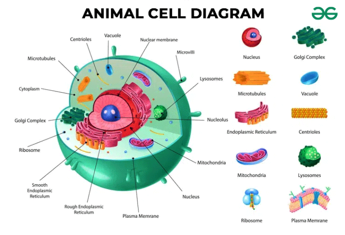
Biology corner animal cell coloring – Hey, fellow Bali beach bum! Let’s dive into the microscopic world, specifically the amazing animal cell – the building block of you, me, and every creature roaming this planet (except plants, they’re a whole other vibe!). Think of it as a tiny, bustling city with specialized departments, all working together in sweet harmony.Animal cells are eukaryotic, meaning they have a membrane-bound nucleus and other organelles – fancy compartments with specific jobs.
This intricate organization allows for efficient cellular processes, keeping everything running smoothly. It’s like a perfectly organized warung, except way smaller and way more complex.
Cell Membrane: The Gatekeeper
The cell membrane is the outer boundary of the animal cell, a selectively permeable barrier that controls what enters and exits. Imagine it as a super-sophisticated bouncer at a trendy Canggu club, only letting in the cool stuff (nutrients, water) and keeping out the riffraff (toxins, unwanted molecules). This membrane is primarily composed of a phospholipid bilayer, a double layer of lipid molecules with hydrophilic (water-loving) heads facing outwards and hydrophobic (water-fearing) tails tucked inside.
Embedded within this bilayer are proteins that act as channels, transporters, and receptors, facilitating the movement of substances across the membrane. This dynamic structure ensures the cell maintains its internal environment, a process crucial for survival. Think of it as maintaining the perfect balance of a delicious smoothie – just the right amount of each ingredient!
Nucleus: The Control Center
The nucleus is the cell’s command center, containing the genetic material (DNA) organized into chromosomes. It’s like the head chef of our cellular warung, dictating what recipes (proteins) to make and when. The DNA holds the blueprints for all cellular activities, and the nucleus regulates gene expression, controlling which proteins are synthesized. The nuclear envelope, a double membrane, surrounds the nucleus, protecting the DNA and regulating the passage of molecules in and out.
Without the nucleus, the cell would be utterly lost, like a chef without a recipe book.
Cytoplasm: The Cellular Workspace, Biology corner animal cell coloring
The cytoplasm is the jelly-like substance filling the cell, excluding the nucleus. It’s where most cellular processes occur. Think of it as the kitchen of our warung – the bustling hub where all the action happens. Various organelles are suspended within the cytoplasm, each performing its specific function. The cytoskeleton, a network of protein filaments, provides structural support and helps with cell movement and intracellular transport.
It’s like the warung’s sturdy framework and efficient delivery system.
Mitochondria: The Powerhouses
These bean-shaped organelles are the energy powerhouses of the cell, generating ATP (adenosine triphosphate), the cell’s primary energy currency. They’re like the warung’s generators, providing the energy needed for all cellular activities. Through cellular respiration, mitochondria break down glucose and other nutrients to produce ATP, fueling everything from muscle contraction to protein synthesis. Without these energetic powerhouses, the cell would simply run out of juice.
Ribosomes: The Protein Factories
Ribosomes are tiny structures responsible for protein synthesis, translating the genetic code from DNA into functional proteins. They are like the warung’s skilled cooks, diligently following the recipes (mRNA) to produce the dishes (proteins) needed for the cell’s various functions. These protein-making machines are found free-floating in the cytoplasm or attached to the endoplasmic reticulum.
Endoplasmic Reticulum (ER): The Manufacturing and Transport System
The ER is a network of interconnected membranes extending throughout the cytoplasm. It’s like the warung’s efficient supply chain and production line. The rough ER, studded with ribosomes, synthesizes proteins, while the smooth ER synthesizes lipids and detoxifies harmful substances. Both work together to ensure smooth and efficient cellular operations.
Golgi Apparatus: The Packaging and Shipping Department
The Golgi apparatus is a stack of flattened sacs that modifies, sorts, and packages proteins and lipids for secretion or transport to other organelles. It’s the warung’s meticulous packaging and shipping department, ensuring everything gets to its destination in perfect condition. This organelle ensures that proteins are correctly folded and delivered to their appropriate locations within or outside the cell.
Biology Corner’s animal cell coloring pages offer a microscopic view of life, a stark contrast to the vibrant macroscopic world. But after meticulously coloring those organelles, you might crave something a bit more…wild! Check out these real animal coloring pages for a change of pace before returning to the intricate details of the cell’s structure. It’s a fun way to appreciate the diversity of life, from the smallest cell to the largest mammal.
Lysosomes: The Waste Recycling Center
Lysosomes are membrane-bound organelles containing digestive enzymes that break down waste materials and cellular debris. They’re like the warung’s efficient waste management system, ensuring everything is recycled or disposed of properly. This process helps maintain cellular cleanliness and prevents the accumulation of harmful substances.
Organelle Focus

Hey, fellow Bali vibes seekers! Let’s dive deep into the amazing world of animal cell organelles. Think of these as the tiny, specialized departments within a bustling cell city, each with its own crucial role to play. We’re gonna explore some key players, getting a real feel for their structure and function – it’s gonna be epic!
Nucleus and Nucleolus
The nucleus is like the cell’s VIP control center, the brain of the operation. It’s a membrane-bound organelle containing the cell’s genetic material, DNA. This DNA is organized into chromosomes, carrying the blueprints for everything the cell does. Think of it as the ultimate instruction manual! Within the nucleus sits the nucleolus, a denser region responsible for producing ribosomes – the tiny protein factories of the cell.
Imagine the nucleolus as a super-efficient workshop churning out essential components for protein synthesis. The nucleus is typically spherical or oval, and its structure includes a double membrane called the nuclear envelope, punctuated by nuclear pores that regulate the transport of molecules in and out.
Mitochondria and Cellular Respiration
Next up, the powerhouses! Mitochondria are the energy generators of the cell, responsible for cellular respiration. This process converts nutrients into ATP (adenosine triphosphate), the cell’s main energy currency. Imagine them as tiny power plants, constantly fueling the cell’s activities. Mitochondria have a unique double-membrane structure: an outer membrane and a highly folded inner membrane called cristae.
These folds significantly increase the surface area for energy production. The more active a cell is, the more mitochondria it typically possesses. For example, muscle cells, requiring a lot of energy for contraction, are packed with mitochondria.
Endoplasmic Reticulum (Rough and Smooth)
The endoplasmic reticulum (ER) is a vast network of interconnected membranes extending throughout the cytoplasm. Think of it as a complex highway system within the cell. There are two main types: rough ER and smooth ER. The rough ER, studded with ribosomes, is the primary site of protein synthesis and modification. The ribosomes synthesize proteins, which are then folded and modified within the rough ER’s lumen before being transported to their final destinations.
The smooth ER, lacking ribosomes, plays a crucial role in lipid synthesis, detoxification, and calcium storage. It’s like the cell’s specialized manufacturing and cleanup crew. Imagine the smooth ER as a dedicated area for lipid production and toxin removal, keeping the cell running smoothly.
| Organelle | Structure | Function | Illustration Description |
|---|---|---|---|
| Nucleus | Spherical or oval; double membrane (nuclear envelope) with pores; contains chromosomes and nucleolus. | Stores genetic material (DNA); controls cell activities; nucleolus produces ribosomes. | A large, round structure with a speckled interior (nucleolus) surrounded by a double membrane showing small pores. |
| Mitochondria | Oval or rod-shaped; double membrane (outer and inner with cristae). | Cellular respiration; ATP production. | Bean-shaped organelles with a folded inner membrane creating numerous compartments within. |
| Rough Endoplasmic Reticulum (RER) | Network of interconnected flattened sacs; studded with ribosomes. | Protein synthesis and modification. | A network of interconnected flattened sacs with numerous small dots (ribosomes) attached to the surface. |
| Smooth Endoplasmic Reticulum (SER) | Network of interconnected tubules; lacks ribosomes. | Lipid synthesis, detoxification, calcium storage. | A network of interconnected tubules, appearing smoother than the RER, without attached ribosomes. |
Cellular Processes & Visualization

Hey, dude! So, we’ve checked out the cool organelles in animal cells, right? Now let’s get into the action – the amazing processes that keep these tiny cities buzzing. Think of it like a super-charged Balinese gamelan orchestra, each instrument (organelle) playing its part to create a harmonious whole. We’re diving into protein synthesis, cell division (mitosis), and how stuff moves in and out of the cell.
Get ready to feel the vibe!
Protein Synthesis
Protein synthesis is like the cell’s ultimate crafting workshop. It’s the process of building proteins, the workhorses of the cell. Imagine a super-detailed blueprint (DNA) in the nucleus, containing the instructions for making a specific protein. This blueprint is transcribed into a messenger RNA (mRNA) molecule, which then heads out to the ribosomes – the protein-building factories. Ribosomes read the mRNA instructions and assemble amino acids, the building blocks of proteins, into a specific sequence, like threading beads onto a string.
The finished protein then folds into a unique 3D shape, ready to perform its specific job. Think of it like a Balinese artisan meticulously crafting a beautiful wood carving, following a precise design to create a unique masterpiece.
Mitosis
Mitosis is cell division – the way cells make copies of themselves. It’s a crucial process for growth, repair, and reproduction in multicellular organisms. Imagine the cell getting ready for a major party, carefully duplicating all its belongings before splitting into two identical daughter cells. The process goes like this: First, the DNA replicates (makes a copy of itself).
Then, the chromosomes condense and align in the center of the cell. Next, the sister chromatids (identical copies of each chromosome) separate and move to opposite ends of the cell. Finally, the cell pinches in the middle, splitting into two genetically identical daughter cells. It’s like a perfectly choreographed Legong dance, with each step precisely executed to create two identical dancers.
Movement of Materials Across the Cell Membrane
The cell membrane is like a selectively permeable gatekeeper, controlling what enters and exits the cell. Imagine it as a bustling marketplace in Ubud, with goods (molecules) flowing in and out. There are different ways materials can cross: Simple diffusion is like molecules casually strolling through the marketplace, moving from an area of high concentration to an area of low concentration.
Facilitated diffusion is like using a shortcut – molecules using protein channels to pass through the membrane more easily. Active transport is like using a porter – molecules are actively pumped across the membrane, requiring energy. Finally, endocytosis is like receiving a large package – the membrane engulfs large particles and brings them into the cell, while exocytosis is the reverse, like shipping out a large order.
Each process is crucial for maintaining the cell’s internal environment, like keeping the marketplace running smoothly.
Question Bank
What are some alternative activities to reinforce learning after coloring?
Build 3D models of cells, create cell-themed board games, or even write short stories from the perspective of an organelle!
How can I adapt this for different age groups?
Younger children can focus on basic organelles and simple coloring, while older students can delve into more complex processes and create detailed diagrams.
Are there any online resources besides Biology Corner that offer similar activities?
Yes! Search for “interactive cell diagrams” or “cell structure games” online to find a wealth of supplementary resources.
What if my students finish early?
Prepare extension activities like researching specific organelles, creating presentations, or designing their own cell-themed artwork.
