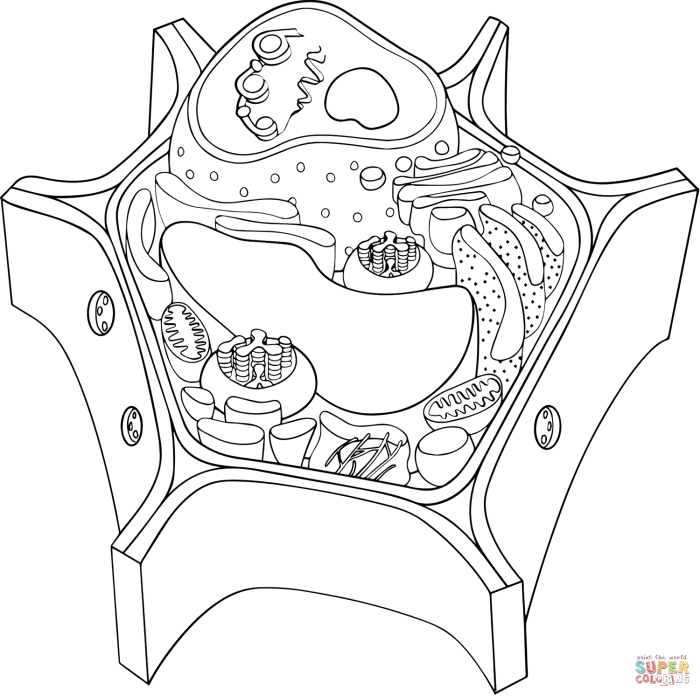
Comparing Staining Techniques for Animal and Plant Cells: Coloring The Cell Animal And Plant

Coloring the cell animal and plant – Staining is a crucial technique in microscopy, allowing visualization of cellular structures that are otherwise transparent. The specific stains and procedures used, however, often differ between animal and plant cells due to variations in their structure and composition.
Staining Procedures for Animal and Plant Cells
The table below summarizes the key differences in staining procedures between animal and plant cells. These differences arise primarily from the presence of a cell wall in plant cells, which acts as a barrier, and the unique organelles found in each cell type.
| Feature | Animal Cell Staining | Plant Cell Staining | Differences |
|---|---|---|---|
| Preparation | Often involves fixation and permeabilization to allow stain penetration. Smearing is common. | May involve sectioning or creating a wet mount. Cell wall permeabilization may be required for some stains. | Plant cell walls require specific treatments for stain penetration. Animal cells are often smeared onto slides, while plant cells are often sectioned or viewed as a whole. |
| Common Stains | Methylene blue, Eosin, Hematoxylin, Giemsa stain | Iodine, Safranin, Toluidine blue, Acetocarmine | Different stains target specific structures present in each cell type. |
| Target Structures | Nucleus, cytoplasm, cell membrane, specific organelles like mitochondria | Cell wall, nucleus, chloroplasts, vacuoles, starch granules | Reflects the unique structural components of each cell type. |
| Mounting | Coverslip placed directly on the stained specimen. | Coverslip placement with mounting media to preserve the specimen. | Mounting media helps maintain the integrity of plant cells and prevents dehydration. |
Reasons for Using Different Stains, Coloring the cell animal and plant
Different stains are used for animal and plant cells because they have varying affinities for specific cellular components. These affinities are based on the chemical properties of the stain and the target structures. For example, some stains bind to negatively charged molecules like DNA and RNA, while others target polysaccharides like those found in plant cell walls.
Organelles Targeted by Staining Methods
Specific stains target distinct organelles within each cell type. For instance, iodine stains starch granules dark blue to black in plant cells, allowing visualization of amyloplasts. In animal cells, methylene blue stains the nucleus a deep blue, highlighting its structure and making it easily distinguishable from the cytoplasm. Eosin, a counterstain often used with hematoxylin, stains the cytoplasm pink.
Safranin stains lignified cell walls red in plant cells. These targeted staining methods allow researchers to study individual organelles and their roles within the cell.
Coloring cell structures of animals and plants can be a great way to learn about their intricate biology. For a more artistic approach, explore detailed animal coloring pages which offer complex designs. This detailed focus on animal anatomy translates well to understanding the nuanced structures within animal and plant cells, furthering appreciation for their complexity.
